


Blood tests feel like mystery boxes: you give a tube, smile nervously, and hope the machine is smarter than your last attempt at assembling flat-pack furniture.
Modern analyzers use precise optics and sensors to decode your blood quickly—backed by WHO laboratory standards for accurate, reliable results.
🩸 Optical Detection Methods: From Colorimetric Reactions to Flow Cytometry
Modern blood analyzers rely on advanced optical systems to read cell counts and chemistry values. They use light, color change, and lasers to turn blood reactions into clear numbers.
These compact devices now deliver lab‑grade accuracy at the bedside. They combine colorimetric tests, photometers, and flow cytometers to monitor health and guide treatment in real time.
1. Colorimetric Reactions and Photometry
Colorimetric tests mix blood with reagents, forming stable colors that match specific analytes such as glucose or hemoglobin. Photometers shine light through the sample and measure absorbance.
- Reagents trigger predictable color changes.
- Light intensity drop relates to analyte level.
- Devices auto‑correct for turbidity and bubbles.
2. Flow Cytometry for Cell Counting
Flow cytometry pushes blood cells in a narrow stream through a laser beam. Detectors read scattered light and fluorescence to classify each single cell.
| Parameter | Measured Feature |
|---|---|
| FSC | Cell size |
| SSC | Cell granularity |
| Fluorescence | Markers on cell surface |
3. Fluorescence Labeling
Fluorescent dyes bind to DNA, RNA, or surface proteins. When excited by lasers, they emit specific colors, helping separate cell types with high precision.
- Improves white cell differentiation.
- Detects immature or abnormal cells.
- Supports immune status monitoring.
4. Optical Design and Error Reduction
Stable light sources, filters, and lenses keep readings accurate over time. Systems monitor drift, reduce background noise, and simplify maintenance for clinical teams.
- Automatic light intensity checks.
- Self‑cleaning cuvettes in some models.
- Built‑in error flags for weak signals.
⚙️ Mechanical and Fluidic Systems: Sampling, Mixing, and Automated Pipetting
Inside a blood analyzer, precise pumps and valves move tiny volumes of blood and reagents. These systems control sampling, dilution, and washing to keep tests stable and fast.
Automated pipetting reduces human error, while smart mixing lowers sample use. This engineering links well with other hospital tools such as an Indoor Rollator or surgical implants that also depend on robust mechanical design.
1. Sample Aspiration and Primary Handling
The device draws blood from tubes using a probe and moves it into internal cups or flow cells. Sensors track volume, clots, and air bubbles.
- Closed aspiration improves safety.
- Level sensors prevent dry aspiration.
- Clot detection avoids blocked channels.
2. Dilution, Mixing, and Reagent Delivery
Accurate dilutions are vital for cell counts and chemistry tests. Mixers use gentle rotation or vibration so cells and reagents blend evenly without damage.
| Step | Function |
|---|---|
| Primary Dilution | Adjusts cell concentration |
| Reagent Add | Starts chemical reactions |
| Mixing | Ensures uniform reaction |
3. Waste Management and Carryover Control
Analyzers flush lines between samples to cut carryover. Waste containers store used reagents and blood safely, following strict infection control rules.
- Smart rinsing sequences.
- Separate hazardous waste paths.
- Sensors alert when tanks are full.
4. Throughput and Workflow Optimization
High‑throughput models coordinate multiple probes and cuvettes. They schedule tasks so different tests run in parallel, reducing turnaround time for busy clinical labs.
🔬 Signal Conversion: Transforming Biochemical Reactions into Electrical Signals
Once light passes through or off the blood sample, detectors convert optical changes into electrical signals. These signals form the base for every clinical result.
Accurate conversion protects patient safety, much like stable support hardware such as WeiMesh Lumbar Titanium Mesh stabilizes the spine during healing.
1. Photodetectors and Analog Signals
Photodiodes and photomultipliers create tiny currents when they receive light. The signal level reflects sample absorbance or fluorescence intensity.
- High sensitivity for low light.
- Low noise electronics.
- Wide dynamic range for many tests.
2. Analog‑to‑Digital Conversion (ADC)
Analog electrical signals pass into ADC chips, which turn them into digital numbers. Resolution and speed affect how fine and fast data can be read.
| ADC Feature | Impact |
|---|---|
| Bit Depth | Detail in small changes |
| Sampling Rate | Ability to follow quick events |
3. Noise Filtering and Signal Stability
Digital filters smooth random spikes from electronics, vibration, or power lines. Stable baselines reduce repeat runs and support consistent clinical decisions.
- Hardware shielding reduces interference.
- Software filters clean raw data.
- Baseline checks catch drift early.
📊 Data Processing Algorithms: From Raw Measurements to Clinically Meaningful Results
Algorithms transform raw counts, light signals, and timing data into final values. These values include cell counts, hemoglobin levels, and complex indices.
Strong processing makes analyzers more than simple readers; they become decision‑ready tools that integrate with lab systems and electronic medical records.
1. Baseline Correction and Curve Fitting
Software first removes background noise and adjusts the zero line. It then fits curves to reaction profiles to interpret concentration and reaction speed.
- Blank correction.
- Drift compensation.
- Nonlinear curve fitting.
2. Classification of Cells and Abnormal Flags
For hematology, algorithms place each cell into size and complexity groups. Pattern rules then mark possible blasts, clumps, or abnormal distributions.
| Cell Group | Typical Use |
|---|---|
| RBC | Detect anemia, polycythemia |
| WBC subsets | Infection, leukemia screening |
| Platelets | Bleeding risk assessment |
3. Reference Ranges, Flags, and Connectivity
The system compares each result with age‑ and sex‑matched ranges. It sets flags when values are critical, then sends structured data to the LIS.
- Automatic delta checks over time.
- Color‑coded result displays.
- HL7 or similar data formats.
🧪 Quality Control and Calibration: Ensuring Accuracy in HUATHENA Blood Analyzers
HUATHENA blood analyzers depend on strict quality control plans. Regular checks, calibrations, and maintenance keep the instruments reliable in busy clinical settings.
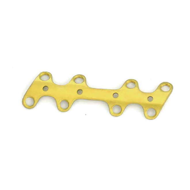
This discipline mirrors the precise engineering seen in devices like the Wedge - shaped Locking Plate - 2 (T), where stable fixation ensures predictable outcomes.
1. Internal and External Quality Control
Labs run control materials with known target values. The analyzer must stay within set ranges before staff release patient reports.
- Multi‑level liquid controls.
- Daily QC charts and rules.
- Automatic lockout when QC fails.
2. Calibration and Reagent Lot Changes
When switching reagent lots or major parts, the system uses calibrators to set new reference points. This keeps results consistent over months and years.
| Event | Required Action |
|---|---|
| New Reagent Lot | Verification or full calibration |
| Optics Service | Recalibrate key channels |
3. Preventive Maintenance and Error Logs
Scheduled cleaning, part replacement, and firmware updates reduce unplanned downtime. Error logs help engineers trace small issues before they affect patients.
- Regular probe and tubing care.
- Filter and lamp checks.
- Remote diagnostics in advanced units.
Conclusion
Modern blood analysis devices combine optics, fluidics, electronics, and smart software to deliver fast, accurate results. Each system step, from sampling to reporting, affects clinical safety.
By understanding these working principles, labs can select the right analyzer, apply strong quality control, and support clinicians with dependable, timely diagnostic data.
Frequently Asked Questions about blood analysis devices
1. How accurate are modern blood analyzers?
Most modern analyzers reach accuracy and precision that meet international standards. When labs follow proper quality control and maintenance, results are stable and suitable for clinical decisions.
2. How long does a typical blood test take on these devices?
Basic complete blood counts often finish in under one minute per sample. High‑throughput systems process many samples in parallel, handling thousands per day.
3. Do small point‑of‑care analyzers work like large lab instruments?
They use similar core principles, such as colorimetry or mini flow cells, but with simpler mechanics and fewer parameters, trading breadth of testing for speed and portability.
4. Why is calibration so important for blood analyzers?
Calibration aligns instrument readings with known standards. Without it, slight drifts in optics, reagents, or electronics can slowly turn into clinically significant errors.
5. How do analyzers protect users from blood exposure?
Closed sampling systems, disposable parts, and secure waste handling limit exposure. Built‑in sensors and error checks further reduce leaks, spills, and contact with hazardous material.